
CUHK
News Centre
CUHK Pioneers the Use of Endoscopic Ultrasonography in Diagnosing Lung Cancers
Lung cancer is the most common malignancy in Hong Kong, with more than 4,000 new cases every year (source: Hong Kong Cancer Registry, Hospital Authority). Treatment options include surgery, radiotherapy and chemotherapy. The choice of treatment relies heavily on accurate staging through tissue sampling of suspicious sites which are often deeply seated. Traditionally, clinicians use computer tomography (CT) scan, bronchoscopy, or even operation under general anaesthesia to evaluate the extent of cancer invasion. These conventional techniques, however, are often traumatic.
Endoscopic ultrasound (EUS) is a flexible endoscope coupled with a small ultrasound device at its tip. Gastroenterologist inserts this endoscope through the patient’s mouth into the oesophagus and stomach. The tip of EUS generates ultrasound wave, which penetrates the wall of the gut and produces visual images of the surrounding internal organs. Because of the close proximity, the image quality is much better than CT scan. Moreover, EUS can safely sample abnormal lymph nodes and lung tissues through the esophagus without the need of general anaesthesia or open surgery.
To assess the diagnostic yield and complication rate of EUS for various suspicious lesions in the chest, 125 patients with abnormal shadows on chest X-ray were recruited from 1998 to 2007 by the Faculty of Medicine at The Chinese University of Hong Kong (CUHK). The diagnosis in about half of these patients remained obscure despite conventional investigations. Using EUS, the causes of abnormal chest X-ray shadows in 83% of the cases were established. Complication rate was very low (<1%) and there was no procedure-related fatality.
“This study suggests that, EUS is an important diagnostic and staging tool for lung cancer patients. It is safe, accurate, and highly tolerable,” said Dr. Larry Lai, the in-charge endoscopist in EUS service and Honorary Clinical Tutor of the Department of Medicine and Therapeutics, Faculty of Medicine, CUHK.
Prince of Wales Hospital, the teaching hospital of CUHK, was the first centre that introduced this novel investigation in Hong Kong, and it is now one of the leading EUS centres in Asia. To date, more than 5,000 patients have undergone EUS at the Prince of Wales Hospital. Apart from lung cancers, EUS has been applied in diagnosing cancers in digestive system, as well as other liver and bile duct problems.

From left: Dr. Yuk Tong LEE, Honorary Associate Professor, Department of Medicine & Therapeutics, CUHK Professor Francis Ka Leung CHAN, Professor of Medicine & Therapeutics, CUHK Dr. Larry H. LAI, the in-charge endoscopist in EUS service and Honorary Clinical Tutor, Department of Medicine & Therapeutics, CUHK Ms. Lee, a lung cancer patient
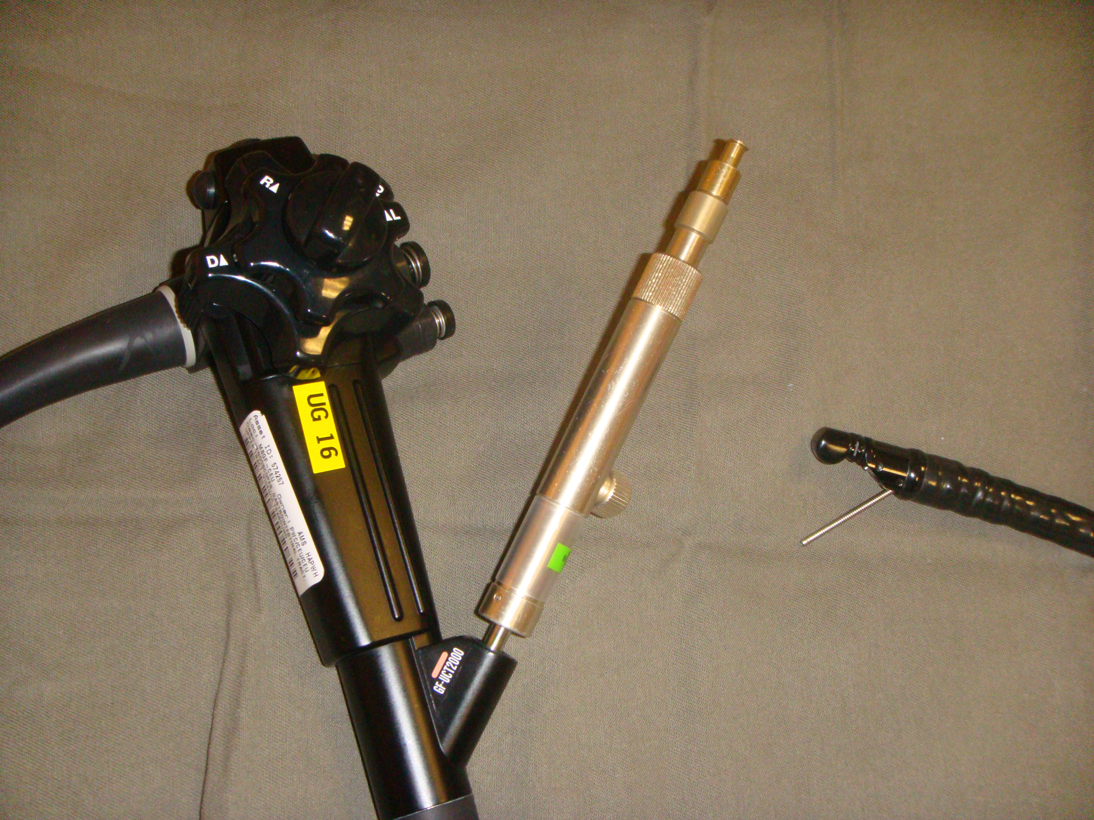
EUS with fine needle aspiration