

CUHK
News Centre
CUHK to Display Award-winning Intestinal Polyp Image Recognition Systemat InnoCarnival 2010
The Chinese University of Hong Kong (CUHK) will take part in the upcoming InnoCarnival 2010 organized by the Innovation and Technology Commission from 6 to 14 November. A total of 19 CUHK technological innovations will be exhibited at Hong Kong Science and Technology Parks to raise public interest in innovative technologies, as well as to educate secondary and primary students. One of the projects displayed will be an intestinal polyp image recognition system, which was the only student project receiving the Best Student Papers Award at the 2009 IEEE International Conference on Robotics and Biomimetics. Three PhD students from the Electronic Engineering Department, Baopu Li, Simon Fan Yichen and Lin Qi, had defeated more than 500 papers from 25 different countries and regions to win this honour.
According to figures released by the Hong Kong Cancer Registry of the Hospital Authorityin December 2009, colorectum cancer is the second most common cancer in Hong Kong. With a total of 4,084 new cases in 2007, it ranks closely behind lung cancer, which has a total of 4,261 recorded cases. The check for intestinal polyp is very important since colorectum cancer is very likely to be cured if diagnosed in an early stage. Capsule endoscopy is one of two methods of intestinal examination.
Resembling a capsule pill in appearance, the capsule endoscope is a medical device for examining the digestive tract. After being swallowed by the patient, it moves through the esophagus and the stomach, finally reaching the intestines. About eight hours after ingestion, it is excreted with the patient’s feces. The camera inside the capsule takes photos of the intestinal conditions, two per second, producing about 50,000 images in one check. It is tiring and time-consuming for physicians to spend an average of two hours on each case to view all the photos taken in order to identify areas with abnormal conditions, such as polyp or bleeding. To solve this problem, Baopu Li, Simon Fan Yichen and Lin Qi invented the intestinal polyp recognition system for capsule endoscopy images under the guidance of Prof. Max Meng.
The system analyzes the hue, saturation and intensity of each photo to sort out images showing polyps according to their colours and shape features and the diagnostic clues employed by physicians. The research team has tested the system with intestinal photos of local patients and found that its accuracy reaches 94.2%, far better than the 50% achieved by similar systems developed in other countries.
The research began in May 2009 and Baopu Li, coordinator of the project, said, ‘This system can greatly reduce the average time required by physicians to review thousands of images captured by a capsule endoscope from more than two hours to less than 30 minutes, thus considerably reducing their workload. We plan to add new functions to the invention to help identify bleeding and ulcer. Practical trials will be carried out in the Prince of Wales Hospital next year and it is hoped that the improved system can provide comprehensive assistance to physicians in intestinal diagnosis.’
At present, the movement of the capsule endoscope can not be controlled by physicians, hampering extensive observation of interested spots along the gastrointestinal (GI) tract, which may result in missed diagnosis. In view of this limitation, the research team led by Prof. Meng is the first to develop the next generation of wireless active capsule endoscope with actuation mechanism and localization system for controlled interactive GI tract examination. The device can be controlled by physicians to examine and stop at any interested spot in the GI tract, and can be guided to move quickly through unimportant areas of the GI tract. The research group adopted a hybrid actuation mechanism which combined an internal actuator and an external magnetic guidance device to help locate the exact position of the capsule by making use of the magnetic field. This new device has improved localization by significantly reducing deviation from 37.5mm to 3mm.
Other key CUHK projects to be displayed at the InnoCarnival 2010 are:
– Ultrasound-guided intervention simulation
– Spatial and appearance correlation of multi-view surveillance videos for in-store customer trace discovery and pro-active selling
About the InnoCarnival 2010
Date: 6 to 14 November 2010
Time: 10am to 7pm
Venue: Hong Kong Science and Technology Parks, Sha Tin (CUHK booth no: C07)
Transport: There will be free shuttle bus service at 15-minute intervals between Hong Kong Science and Technology Parks, and the MTR Shatin and University Station respectively (frequency of the bus will depend on number of passengers.)
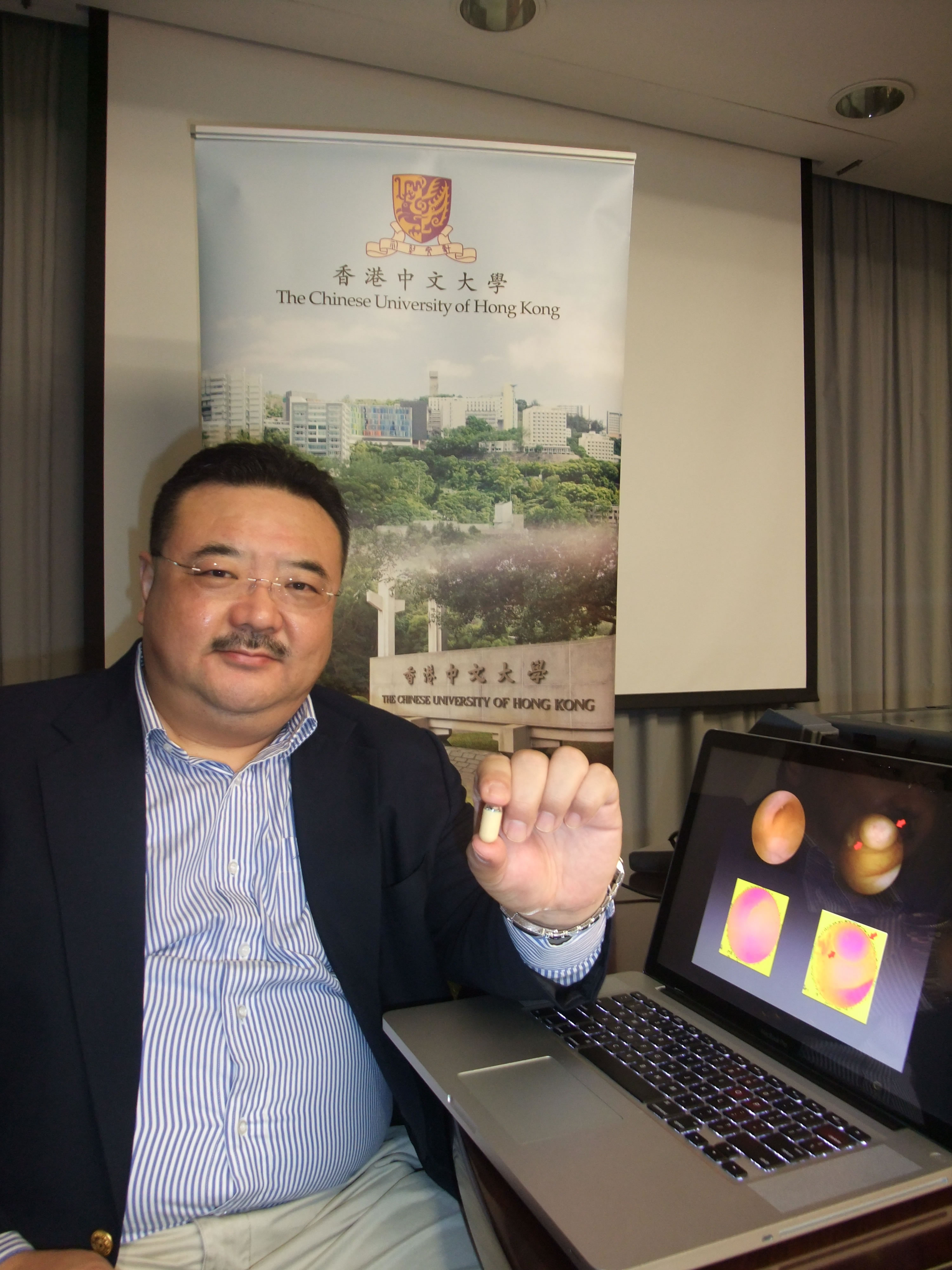
Prof. Max Meng and his research team at the Electronic Engineering Department of CUHK has greatly enhanced the function of capsule endoscope
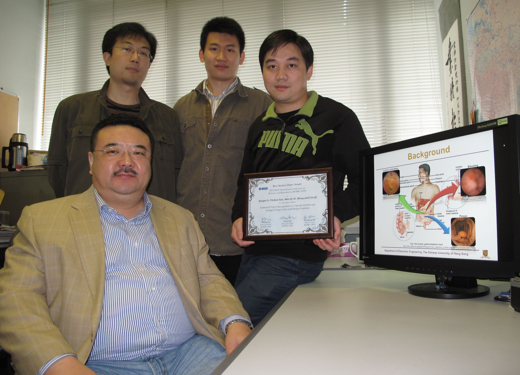
Prof. Max Meng and his PhD students (from left): Lin Qi, Simon Yichen Fan, and Baopu Li
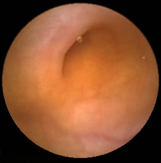
Normal intestinal image taken by a capsule endoscope
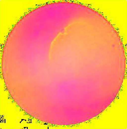
Normal intestinal image after having its hue, saturation and intensity analysed by the system
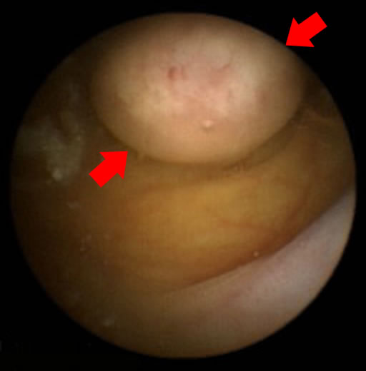
Intestinal polyp image taken by a capsule endoscope
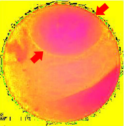
Intestinal polyp image after having its hue, saturation and intensity analysed by the system