

CUHK
News Centre
A CUHK-led international team develops the world’s first AI model using fundus photographs alone to detect Alzheimer’s disease
The retina is an extension of the central nervous system; thus, the eye is therefore a window that can show degenerative changes in the blood vessels and nerves of the brain. An international team led by The Chinese University of Hong Kong (CUHK)’s Faculty of Medicine (CU Medicine) has successfully developed the world’s first artificial intelligence (AI) model that can detect Alzheimer’s disease solely through fundus photographs or images of the retina. The model is more than 80% accurate after validation.
Considering fundus photography is widely accessible, non-invasive and cost-effective, this novel AI model incorporated with fundus photography is expected to become an important tool for screening people at high risk of Alzheimer’s disease in the community. Details have been published in The Lancet Digital Health under the international journal The Lancet.
Current methods to detect early Alzheimer’s disease are limited
In Hong Kong, 1 in 10 people aged 70 or above suffers from dementia, with more than half of those cases attributed to Alzheimer’s disease. Alzheimer’s disease is associated with an excessive accumulation of abnormal amyloid plaque and neurofibrillary tangles in the brains, leading to the death of brain cells and resulting in progressive cognitive decline.
Dr Lisa Au Wing-chi, Clinical Professional Consultant of the Division of Neurology in CU Medicine’s Department of Medicine and Therapeutics, said, “Memory complaints are common among middle-aged and elderly people, and often considered a sign of Alzheimer’s disease. It is sometimes difficult to make an accurate diagnosis of Alzheimer’s disease based on cognitive tests and structural brain imaging. However, methods to detect Alzheimer’s pathology, such as an amyloid-PET scan or testing of cerebrospinal fluid collected via lumber puncture, are invasive and less accessible.”
To address the current clinical gap, CU Medicine has led a number of medical centres and institutions from Singapore, the United Kingdom and the United States to successfully develop an AI model using state-of-the art technologies which can detect Alzheimer’s disease using fundus photographs alone.
The retina is a window to study disorders of the central nervous system
Professor Clement Tham Chee-yung, S.H. Ho Professor of Ophthalmology and Visual Sciences and Chairman of CU Medicine’s Department of Ophthalmology and Visual Sciences, explained, “The retina is an extension of the brain in terms of embryology, anatomy and physiology. In the entire central nervous system, only the blood vessels and nerves in the retina allow direct visualisation and analysis. Hence, it has long been considered a window to study disorders in the central nervous system. Through non-invasive fundus photography, we can detect a range of changes in the blood vessels and nerves of the retina that are associated with Alzheimer’s disease.”
The team developed and validated their AI model using nearly 13,000 fundus photographs from 648 Alzheimer’s disease patients (included patients from the Prince of Wales Hospital) and 3,240 cognitively normal subjects. Upon validation, the model showed 84% accuracy, 93% sensitivity and 82% specificity in detecting Alzheimer’s disease. In the multi-ethnic, multi-country datasets, the AI model achieved accuracies ranging from 80% to 92%.
Accessibility, non-invasiveness and high cost-effectiveness of the AI model using fundus photography help detection of Alzheimer’s cases both in clinic and the community
Professor Vincent Mok Chung-tong, Mok Hing Yiu Professor of Medicine and Director of the Therese Pei Fong Chow Research Centre for Prevention of Dementia at CU Medicine, remarked, “In addition to its accessibility and non-invasiveness, the accuracy of the new AI model is comparable to imaging tests such as magnetic resonance imaging (MRI). It shows potential to become not only a diagnostic test in clinics, but also a screening tool for Alzheimer’s disease in community settings. In the future, we hope to validate its efficacy in identifying high-risk cases of the disease hidden in the community, so that various preventive treatments such as anti-amyloid drugs can be initiated early to slow down cognitive decline and brain damage.”
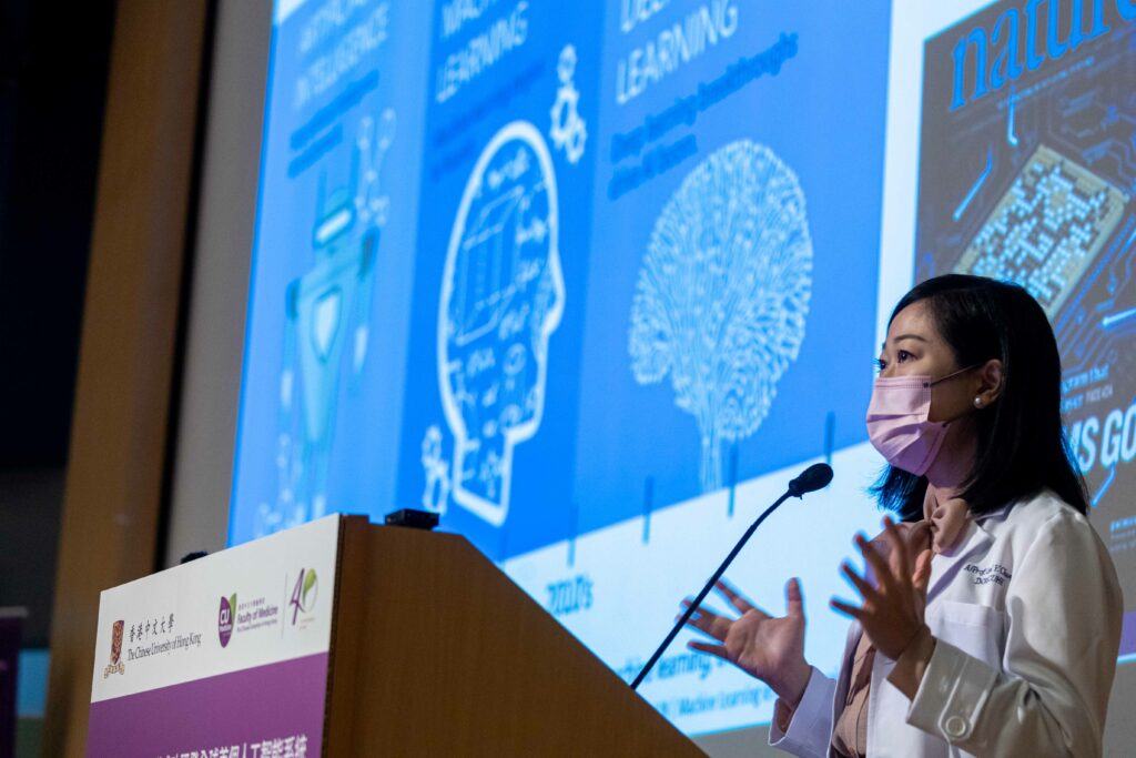
Dr Carol Cheung Yim-lui, Associate Professor in the Department of Ophthalmology and Visual Sciences at CU Medicine, added, “In addition to applying novel AI technologies in the model, we also tested it in different scenarios. Notably, our AI model retained a robust ability to differentiate between subjects with and without Alzheimer’s disease, even in the presence of concomitant eye diseases like macular degeneration and glaucoma which are common in city-dwellers and the older population. This further supports the concept that our AI analysis of fundus photographs is an excellent tool for the detection of the memory-depriving Alzheimer’s disease. To move this research towards clinical application, we are developing an integrated, AI-based platform to combine information from both blood vessels and nerves of the retina captured by fundus photography and optical coherence tomography for detection of Alzheimer’s disease. Our findings should provide more evidence to move AI from code to the real world.”
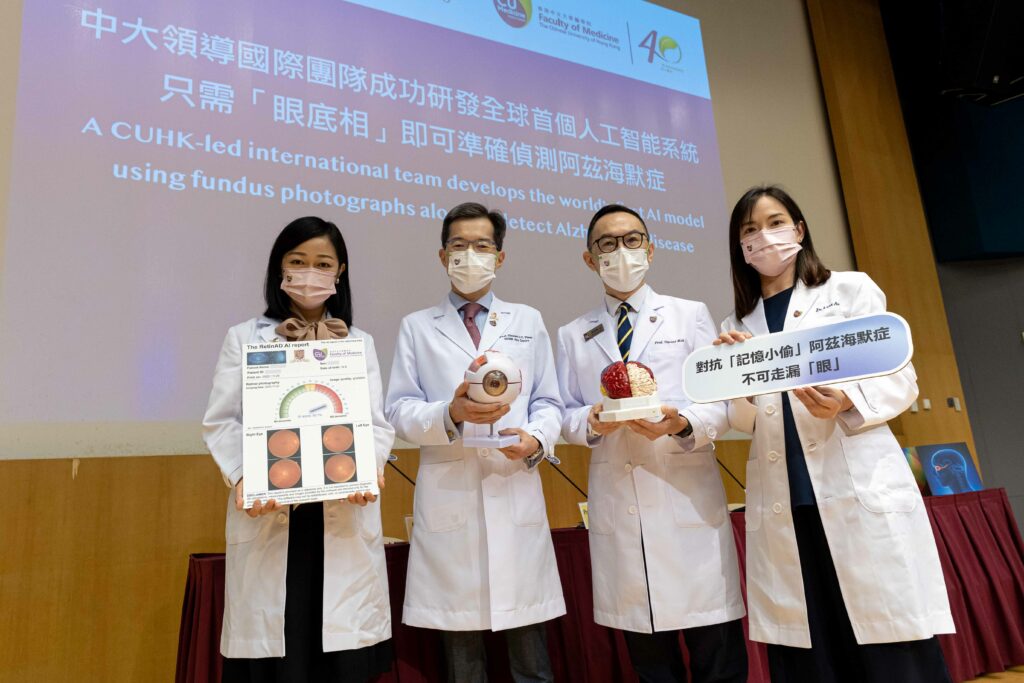


An international team led by CU Medicine has successfully developed the world’s first artificial intelligence (AI) model that can detect Alzheimer’s disease solely through fundus photographs. The model is more than 80% accurate after validation. Considering fundus photography is widely accessible, non-invasive and cost-effective, this novel AI model is expected to become an important tool for screening people at high risk of Alzheimer’s disease in the community.
(From left) Dr Carol Cheung, Associate Professor in the Department of Ophthalmology and Visual Sciences; Professor Clement Tham, S.H. Ho Professor of Ophthalmology and Visual Sciences and Chairman of the Department of Ophthalmology and Visual Sciences; Professor Vincent Mok, Mok Hing Yiu Professor of Medicine and Director of the Therese Pei Fong Chow Research Centre for Prevention of Dementia; Dr Lisa Au, Clinical Professional Consultant of the Division of Neurology in Department of Medicine and Therapeutics; at CU Medicine.

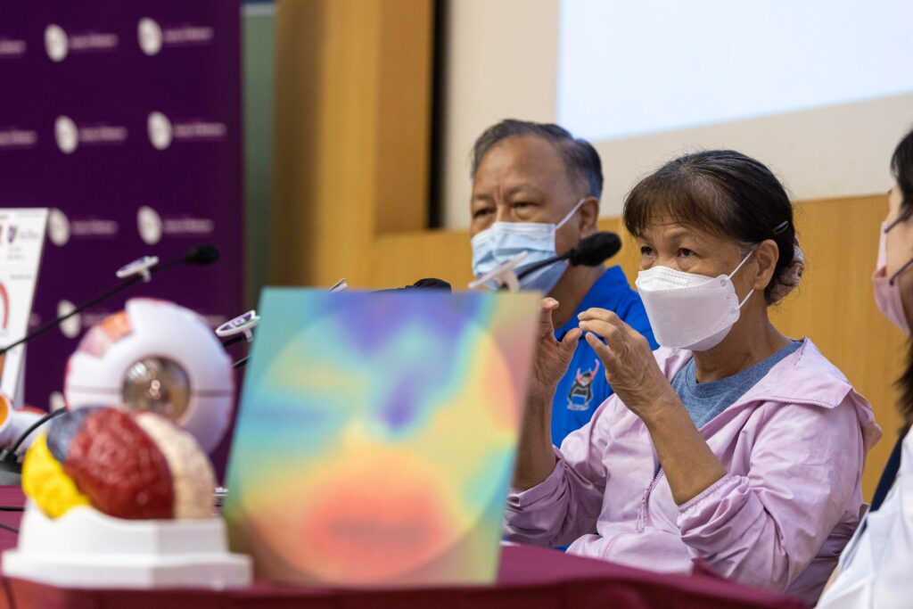
Alzheimer’s disease patient Mrs Chan (centre) says that the process of taking fundus photographs was simple and she did not feel any discomfort.

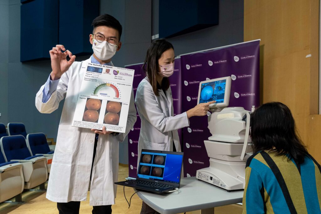
The research team introduces that the process of taking fundus photographs is quick and convenient. The AI system takes about one minute to calculate the “risk assessment score” to detect the risk of Alzheimer’s disease.

Professor Vincent Mok points out that the new AI model is accessible, non-invasive and accurate, showing its potential to become a diagnostic test in clinics for Alzheimer’s disease in clinic settings.
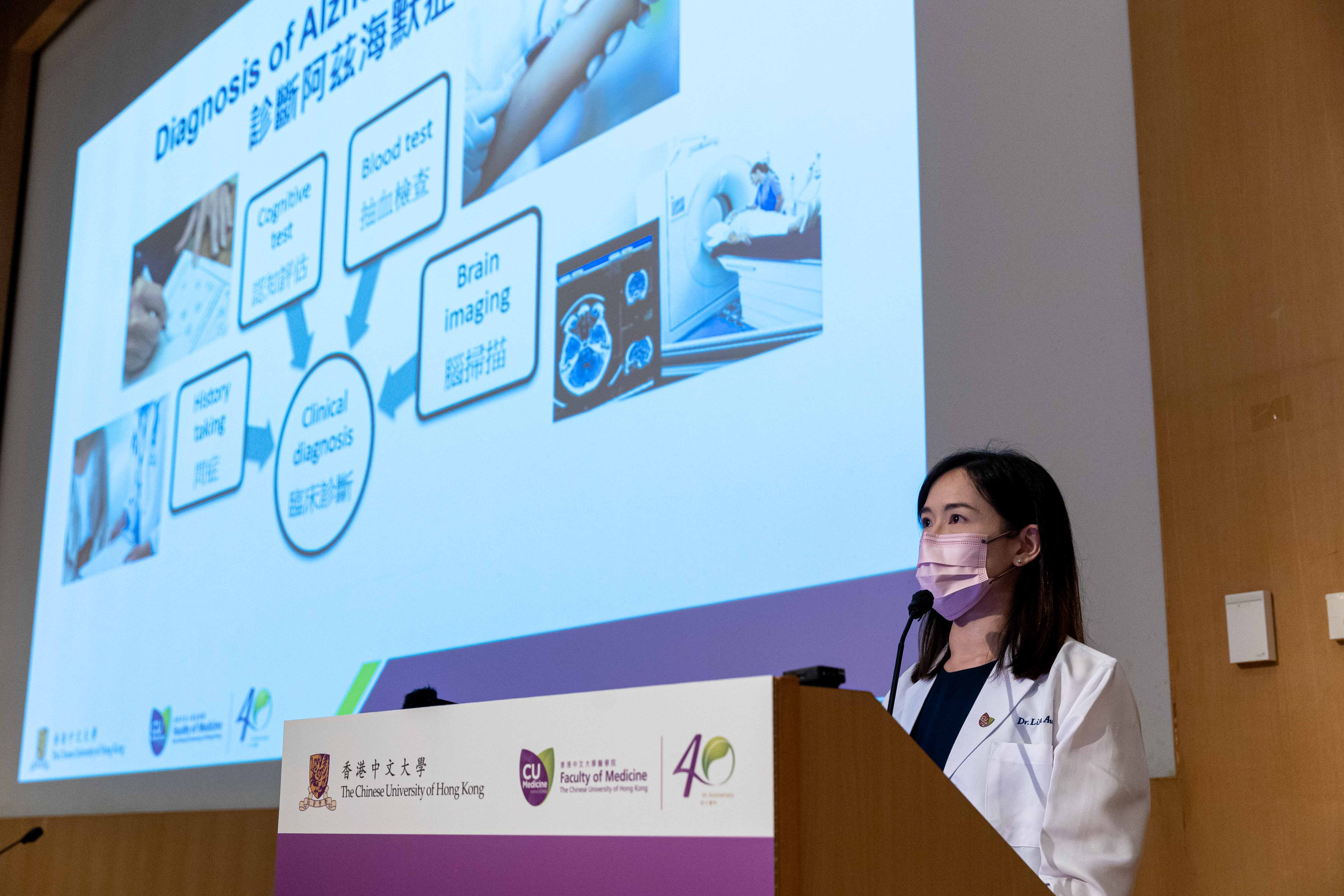
Dr Lisa Au states that it is sometimes difficult to make an accurate diagnosis of Alzheimer’s disease based on cognitive tests and structural brain imaging.
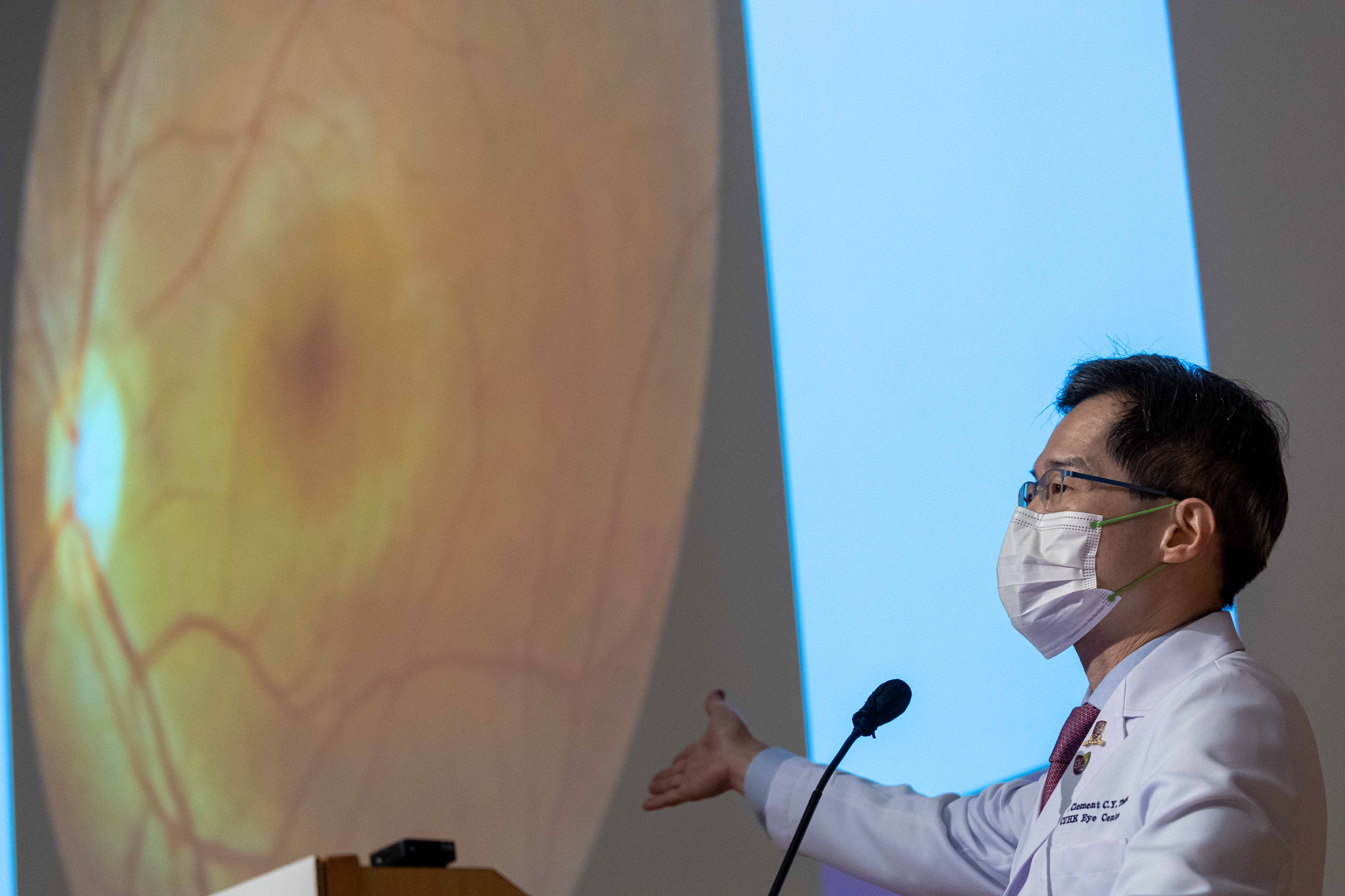
Professor Clement Tham explains that in the entire central nervous system, only the blood vessels and nerves in the retina allow direct visualisation and analysis. Through non-invasive fundus photography, a range of changes that are associated with Alzheimer’s disease can be detected.
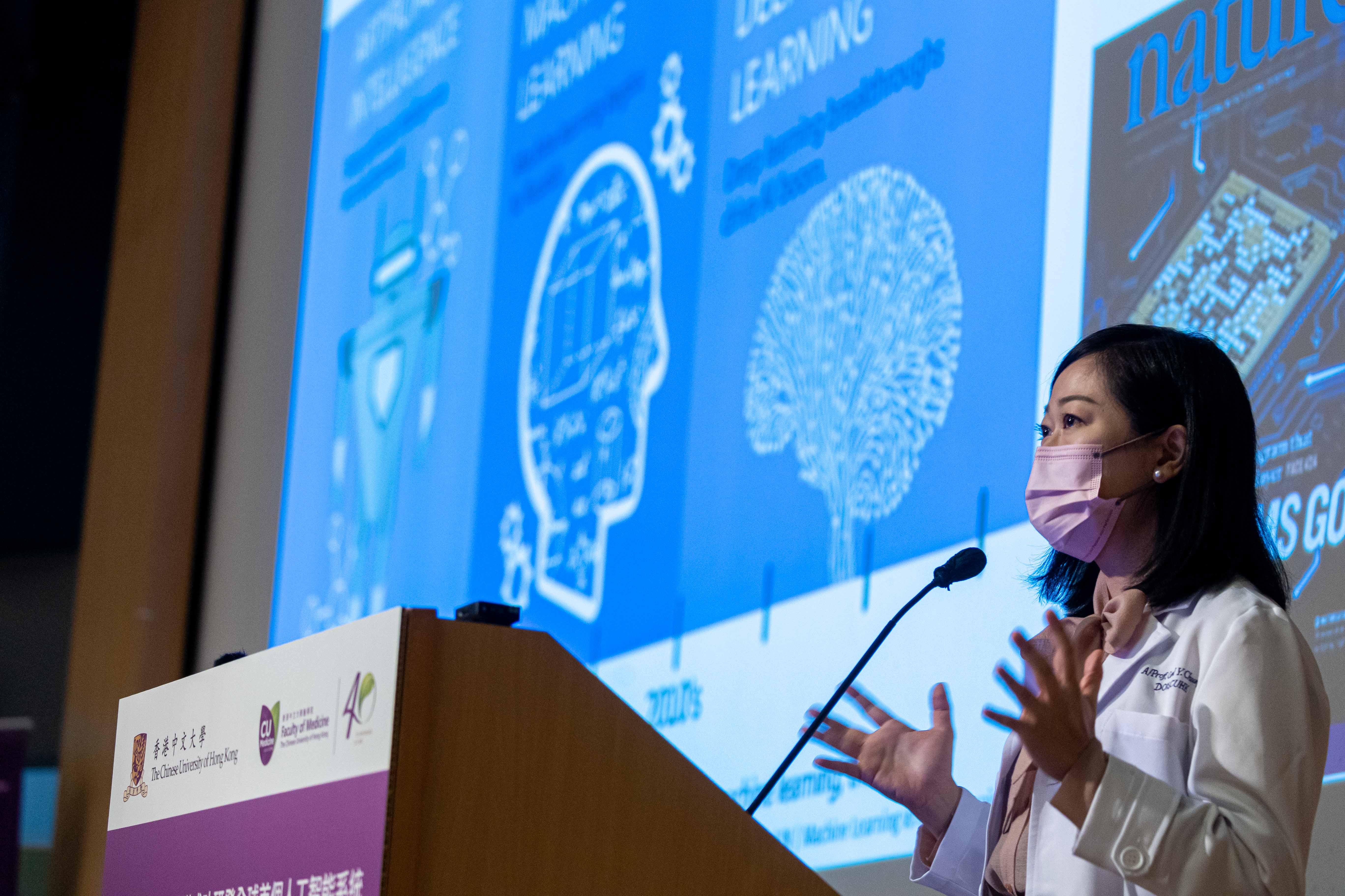
Dr Carol Cheung states that the novel AI model has been tested in different scenarios. Notably, the model retained a robust ability to differentiate between subjects with and without Alzheimer’s disease, even in the presence of concomitant eye diseases like macular degeneration and glaucoma which are common in city-dwellers and the older population.