

CUHK
News Centre
Intestinal Polyp Image Recognition System Invented by CUHK PhD Students Won IEEE Award
Three PhD students of the Electronic Engineering Department at The Chinese University of Hong Kong (CUHK) have developed a new image recognition system that can handle huge amounts of images captured by capsule endoscopy. This system, with more than 90% accuracy in distinguishing intestinal polyp images from ones showing normal regions, has greatly shortened the time required by physicians to sort images with their own eyes. Defeating more than 500 papers from 25 different countries and regions, this invention won the Best Student Papers Award, the only student award, at the 2009 IEEE International Conference on Robotics and Biomimetics last month.
According to statistics released in December 2009 by the Hong Kong Cancer Registry of Hospital Authority, colorectum cancer is the second most common cancer in Hong Kong. With a total of 4,084 new cases in 2007, it ranks closely behind lung cancer, which has a total of 4,261 recorded cases. The check for intestinal polyp is very important since colorectum cancer is very likely to be cured if diagnosed in an early stage. Capsule endoscopy is one of two methods of intestinal examination.
Resembling a capsule pill in appearance, the capsule endoscope is a medical device for examining the digestive tract. After being swallowed by a patient, it moves through the esophagus, stomach, and finally reaching the intestines. It is excreted with the patient’s feces about eight hours after ingestion. The camera inside the capsule takes photos of the intestinal conditions, two per second, producing about 50,000 images in one check. It would be tiring and time-consuming for physicians to spend an average of two hours on each case to view all the photos taken in order to identify areas with abnormal conditions, such as polyp or bleeding. To solve this problem, three PhD students, Baopu Li, Simon Yichen Fan and Lin Qi of the Electronic Engineering Department, invented the ‘Intestinal Polyp Recognition System for Capsule Endoscopy Images’ under the guidance of Prof. Max Meng of the department.
The system analyses the hue, saturation and intensity of each photo to sort out images showing polyps according to their colours and shape features and the diagnostic clues employed by physicians. The research team has tested the system with intestinal photos of local patients and found that its accuracy reaches 94.2%, far better than the 50% achieved by similar systems developed in other countries.
The research began in May 2009 and Baopu Li, coordinator of the project, said, ‘This system can greatly reduce the average time required by physicians to review thousands of images captured by a capsule endoscope from more than two hours to less than half an hour, thus considerably reducing their workload. We plan to add new functions to the invention to help identify bleeding and ulcer. Practical trials will be carried out in the Prince of Wales Hospital next year and it is hoped that the improved system can provide comprehensive assistance to physicians in intestinal diagnosis.’
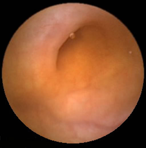
Normal intestinal image taken by a capsule endoscope
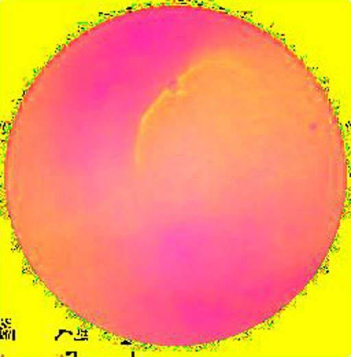
Normal intestinal image after having its hue, saturation and intensity analysed by the system
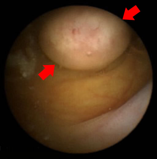
Intestinal polyp image taken by a capsule endoscope
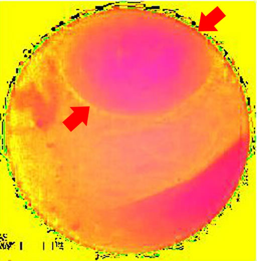
Intestinal polyp image after having its hue, saturation and intensity analysed by the system
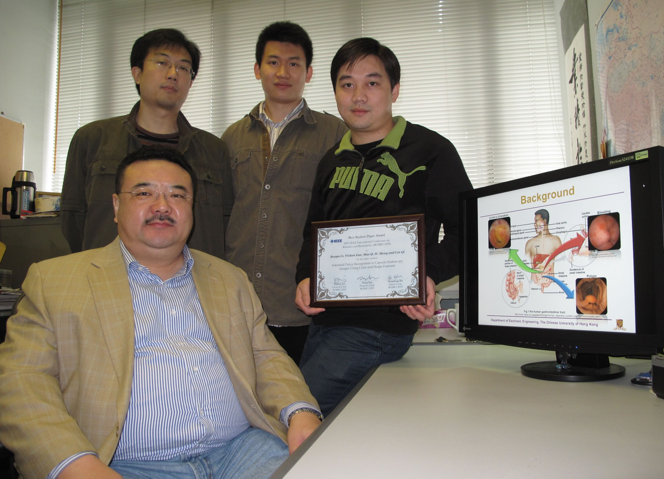
Prof. Max Meng (front) and his PhD students (from left): Lin Qi, Simon Yichen Fan, and Baopu Li