

CUHK
News Centre
Smart Hong Kong City Development SeriesCUHK Faculty of Engineering Develops Ultrafast Microscope to Help Tackle Glaucoma and Other Neurological Diseases
A microscope that may help the study of neurological diseases has been developed at The Chinese University of Hong Kong (CUHK). A team led by Prof. Shih-Chi Chen, Associate Professor in the Department of Mechanical and Automation Engineering at CUHK, has recently developed the first digital holography-based (DH) two-photon excitation (TPE) microscope to generate simultaneous video-rate fluorescent imaging and multi-point optical stimulation. This allows the tracking of nerve cells activities and thus may help the study of neurological diseases, such as glaucoma, a very common eye disease.
By scanning the retinal ganglion cells using the DH-TPE microscope, scientist can understand the molecular mechanisms of optic nerve degeneration in glaucoma and find ways to tackle it. Prof. Chen’s team is now working on this project with Prof. Christopher Kai Shun Leung, Professor, Department of Ophthalmology and Visual Sciences at the Faculty of Medicine at CUHK, and the Hong Kong Eye Hospital.
3-D imaging tracks inter-cells activities which helps the study of glaucoma
The DH-TPE microscope is powered by a digital micromirror device (DMD), a chip used in our everyday projector that contains millions of micromirrors switching at tens of kilohertz speed. By controlling the amplitude and phase of the input laser via binary holograms and the fast-switching micromirrors, the laser beam can be split into up to 20 focal points for simultaneous optical stimulation and real-time fluorescent imaging. Each focus can be independently controlled to scan along arbitrarily defined paths or surfaces at 22.7 kHz. More importantly, the DMD-scanner is also an ultrafast beam shaper, and by superposing the scanning and wavefront-correction holograms, the point spread function can be engineered or even shaped into other novel beam modes to achieve efficient 3-D imaging. Compared to state-of-the-art commercial TPE microscopes, the DH-TPE microscope presents a suite of distinctive imaging functionalities that have never been realised in the past, including (1) random-access imaging, (2) multi-plane imaging, (3) 3-D programmable imaging plane, (4) point-specific wavefront correction, and (5) simultaneous video-rate fluorescent imaging and multi-point optical stimulation.
Through funding support from the Innovation and Technology Commission (ITC), Prof. Chen’s team is setting up the DH-TPE microscope at the Hong Kong Eye Hospital, collaborating with a team led by Prof. Christopher Kai Shun Leung. The two teams are working together to exploit the unique capability of the DH-TPE microscope to study and understand the basic mechanisms of a few important diseases.
For example, one objective is to study metabolic dysfunction in the retinal ganglion cells via in vivo two-photon imaging to study the molecular mechanisms of optic nerve degeneration in glaucoma. Glaucoma, characterised by progressive loss of retinal ganglion cells, is the leading cause of irreversible blindness worldwide with a significant social and economic burden. The reduced form of nicotinamide adenine dinucleotide (NADH) is an intrinsic fluorophore and a co-factor in major metabolic pathways for energy production. The levels of NADH decrease in the retinal ganglion cell following optic nerve injury and the imaging of NADH fluorescence intensity can serve as a non-invasive indicator of retinal ganglion cell death in glaucoma. The unique design of the DH-TPE microscope allows point-specific wave-front correction of ocular aberration that will facilitate non-invasive, label-free imaging of the retina. The investigation of metabolic dysfunction of retinal ganglion cells will provide mechanistic insights into the development of neuroprotective and neuroregenerative therapies for patients with glaucoma and non-glaucomatous optic neuropathies.
CUHK Engineering Series: Smart Hong Kong City Development
CUHK is one of the pioneers in the research on artificial intelligence (AI) and deep learning. It has already received top awards in international challenges for face recognition and visual recognition. The CUHK Multimedia Laboratory was named as one of the pioneers in AI research, the only institution in Asia to receive this recognition, alongside the University of California, Berkeley, Massachusetts Institute of Technology, Stanford University and the University of Oxford. Different research teams in the Departments of Information Engineering, Electronic Engineering, Systems Engineering and Engineering Management, and Computer Science and Engineering at the Faculty of Engineering are also dedicated to the application of AI and deep learning in various areas, such as smart living, smart mobility and smart environment, in order to build Hong Kong into a world class smart city. In the future, the Faculty will launch more innovative projects that make use of AI to improve the quality of human life and share it with the public through the ‘Smart Hong Kong City Development’ Series.
About The Chinese University of Hong Kong
Founded in 1963, CUHK is a leading comprehensive research university with a global reputation. Located in the heart of Asia, CUHK has a vision and a mission to combine tradition with modernity, and to bring together China and the West. Under the University’s unique collegiate system, the programmes and activities offered by its nine colleges complement the formal curricula by delivering whole-person education and pastoral care. The University has eight faculties: Arts, Business Administration, Education, Engineering, Law, Medicine, Science, and Social Science. Together with the Graduate School, the University offers over 300 undergraduate and postgraduate programmes. All faculties are actively engaged in research in a wide range of disciplines, with an array of research institutes and research centres specialising in interdisciplinary research of the highest quality.
CUHK is recognised as the most innovative university in Hong Kong and ranks 22nd in the Asia-Pacific region by the latest ‘Reuters: Asia Pacific’s Most Innovative Universities’ listing. The University currently has more than 800 granted patents in different jurisdictions worldwide. Some of these patents have been licensed to relevant industries that help bring these innovations to the market to benefit society. In academic year 2016-17, CUHK has received 132 granted patents and filed 223 patent applications for inventions developed in the areas of medical technology, biotechnology, information technology, telecommunications, and materials science.
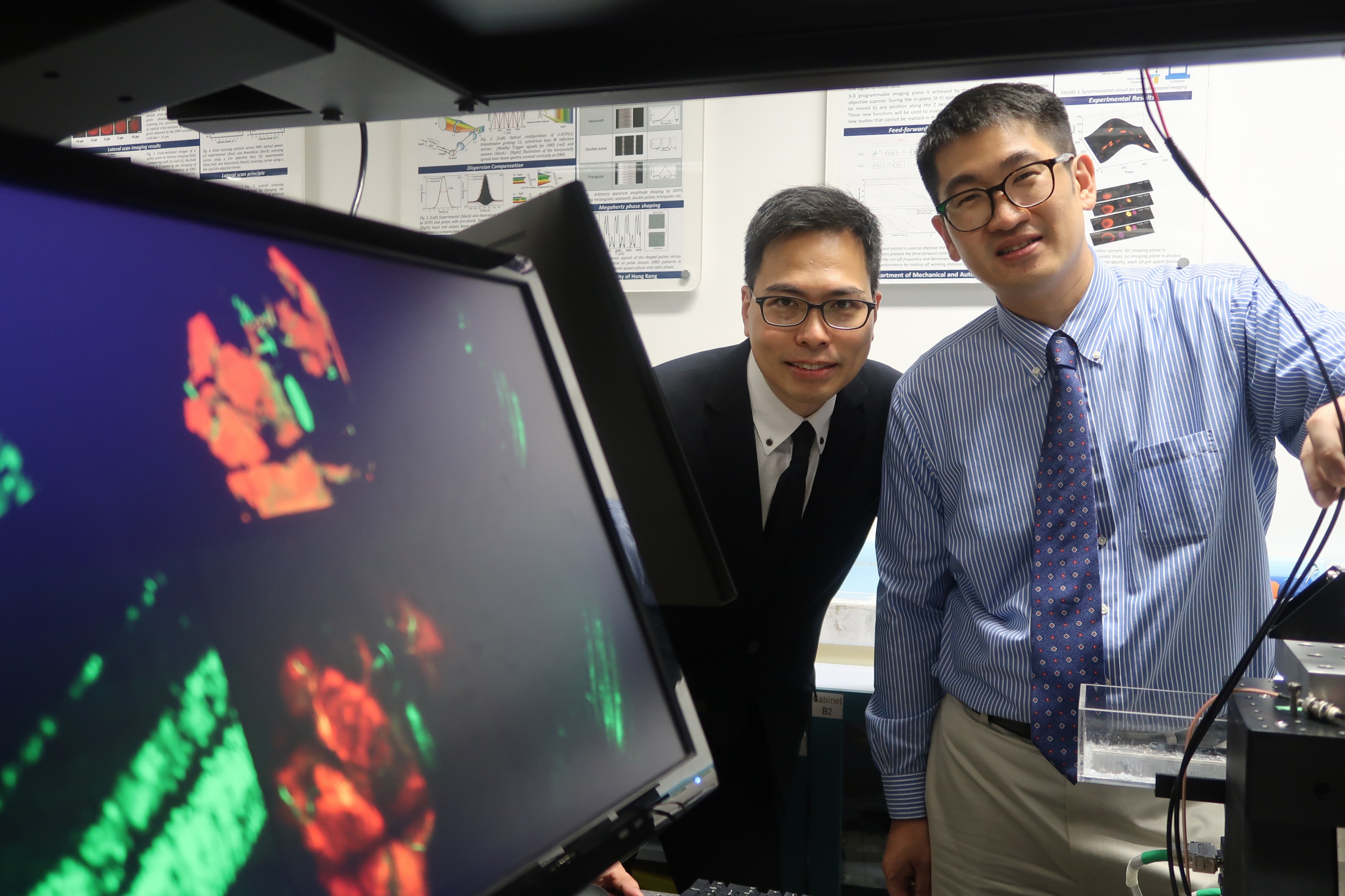
Using the DH-TPE microscope, a new invention of CUHK, Prof. Shih-Chi Chen from the Faculty of Engineering and Prof. Christopher Kai Shun Leung from the Faculty of Medicine scan retinal ganglion cells of living animals to find out the molecular mechanisms of optic nerve degeneration in glaucoma.
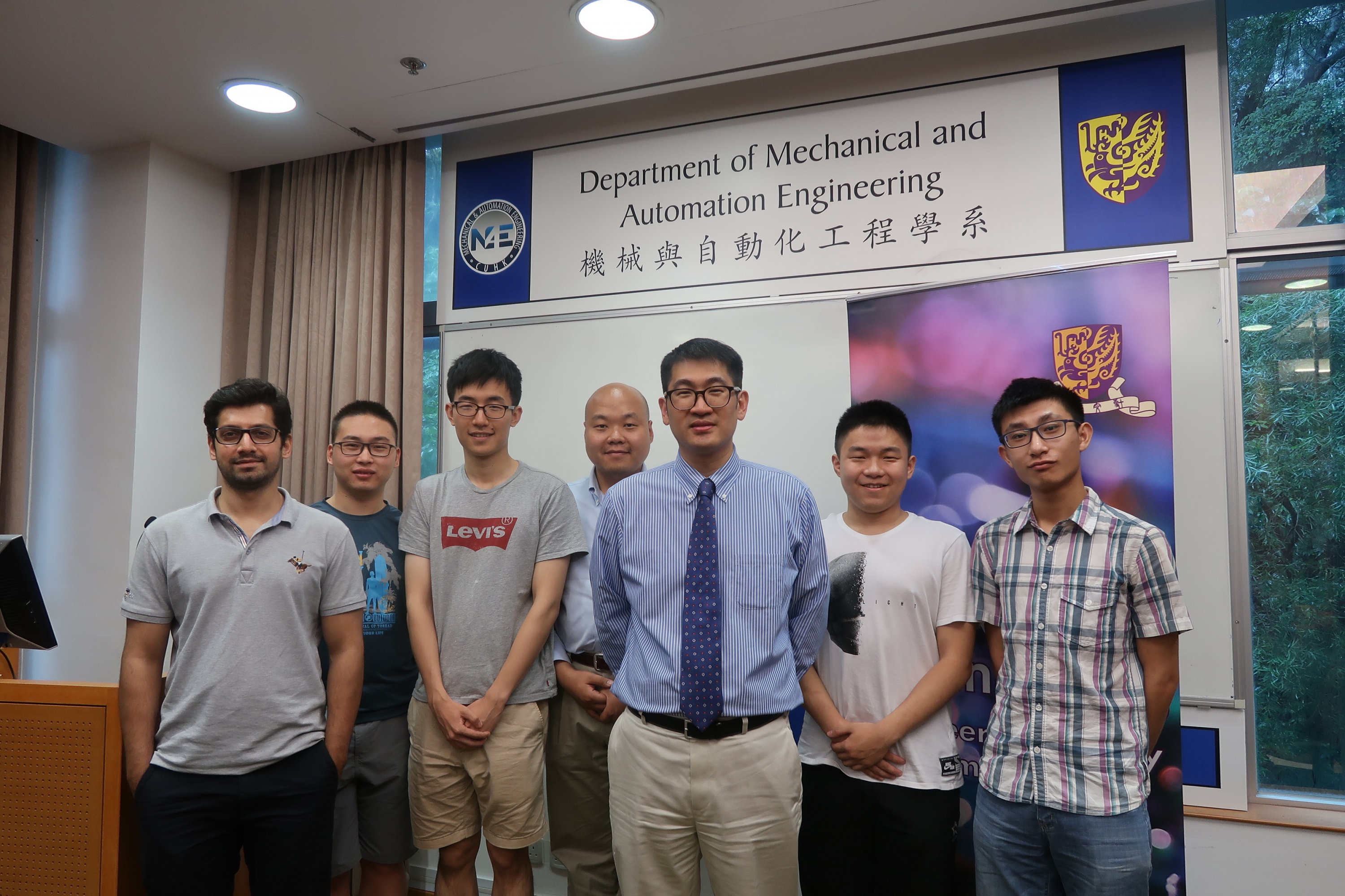
The research team of Prof. Shih-Chi Chen (3rd right).
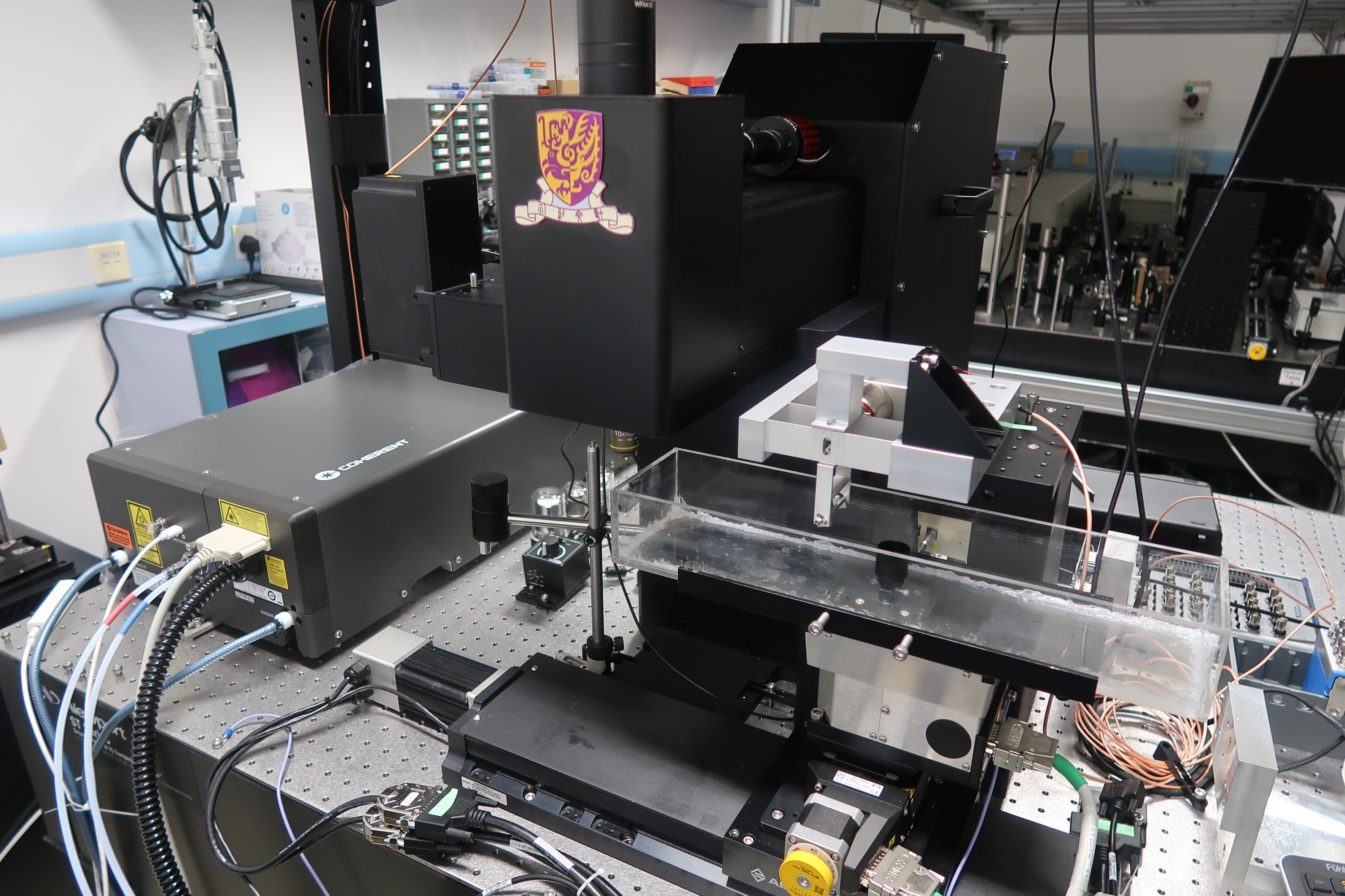
DH-TPE microscope is powered by a digital micromirror device. It can generate simultaneous video-rate fluorescent images, which allow the tracking of nerve cells activities.